Freezer Fracture
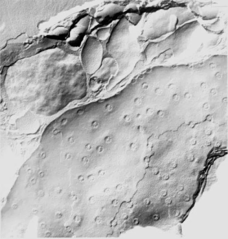
Freeze fracturing is a process where a frozen specimen is cracked to reveal a plane through the tissue. The fracture occurs along weak hydrophobic planes such as membranes or surfaces of organelles. The technique is extremely powerful when applied to the study of membrane structure and organisation.
Once fractured, replicas of fractured surfaces are prepared and separated from the tissue leaving a thin metal (eg platinum-carbon) replica to examine in a transmission electron microscope (TEM). Replicas are uniquely suited for the study of membrane structure, notably the internal aspects of the lipid bilayer, thus providing a three-dimensional representation of the tissue and cellular landscape. This technique is very technically demanding and slow with the fragile replica requiring careful cleaning before being collected on a TEM grid for viewing.
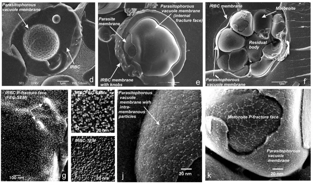
Instrument advances now allow fully hydrated fracture faces to be directly imaged at high resolution in a cryogenic field emission gun scanning electron microscope. The technique relies on vitrification of tissues by high pressure freezing, fracturing and ion beam coating at high vacuum conditions and cryogenic temperatures.
Examples of use
We have applied freeze fracture in numerous ways to assist in research.
Freeze Fracture TEM
Freeze fracture allowed the effects of gene regulation on membrane organisation to be studied and the functional changes which stabilise membranes against freeze-induced injury to be understood. In Arabidopsis, transcription factors have been identified that control the expression and regulation of cold-induced genes that increase plant freezing tolerance – a process known as cold acclimation.
Freeze fracture cryogenic scanning electron microscopy (cryo-SEM)
High resolution freeze fracture cryo SEM of infected red cell membrane allows specialised membrane structures to be studied. These structures known as ‘knobs’ appear on the surface of malaria infected erythrocytes and are involved in cytoadherence.
Equipment available
- Leica microsystems ACE900.


