This is the result of a truly multi-disciplinary effort, finding the compromise between engineering a perfect solution and adapting to available clinical data – a journey built on pillars of mutual trust and recognition among the research team and fueled by the energy of the perfect duet, Joao and Otto.
Pablo Lamata, Professor at the School of Biomedical Engineering & Imaging Sciences
21 January 2025
Researchers develop non-invasive method to assess diastolic function via pressure quantification in the heart
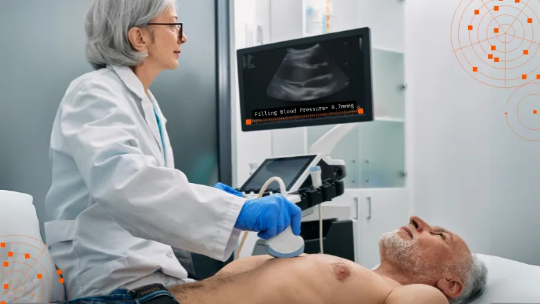
A team of researchers from the School of Biomedical Engineering & Imaging Sciences, Rikhospitalet in Norway and partners in Japan and France have developed a groundbreaking non-invasive method to measure how the heart fills with blood during diastole. Their innovation aims to improve the diagnosis and treatment of heart failure and related conditions.

The researchers, led by Professors Pablo Lamata and Otto Smiseth, used conventional echocardiography, a widely available ultrasound imaging technique, to estimate the blood pressure inside the heart’s main chamber during its relaxation phase. The study, published in the European Heart Journal, combines imaging data with mathematical models to provide a detailed picture of diastolic function without the need for invasive procedures.
This advancement addresses a long-standing gap in cardiology: while tools like blood pressure cuffs measure the force of the heart’s pumping, assessing the heart’s ability to fill with blood has been much more challenging. By estimating pressures inside the heart during diastole, the new method offers critical insights into conditions such as heart failure with preserved ejection fraction (HFpEF), where the heart struggles to fill effectively despite normal pumping function.
The key innovation involves analyzing the pressures generated during different phases of the heart’s diastolic filling. This includes the minimum pressure, which reflects how well the heart relaxes, and the end-diastolic pressure, which indicates the heart’s readiness to pump blood. These measurements were validated against invasive techniques and showed a high degree of accuracy.
"Echocardiography is already a mainstay of clinical practice,” said Dr Joao Fernandes, who spearheaded the engineering work, “and this method builds on routinely collected data, making it a promising addition to heart failure diagnostics.”
The researchers hope this non-invasive approach will improve the characterization of heart failure and assist in tailoring treatments. For instance, in patients with pulmonary congestion, understanding the pressures inside the heart could help refine therapy to reduce symptoms and improve outcomes. Future studies will aim to test the method in larger, more diverse populations and explore its potential to guide personalized care for heart disease patients.
The outcome of this paper addresses one of the holy grails in cardiology: the non-invasive quantification of filling pressure. We are working on the real potential for clinical translation and future clinical applications, and I want to emphasize that the 5-year character-building journey that has led to our cornerstone publication - is only just beginning.
Dr Joao Fernandes, Research Associate at the School of Biomedical Engineering & Imaging Sciences


