Transmission electron microscopes (TEMs) are powerful analytical tools for investigating very small structures. TEMs can show all the structures of a tissue and compartmentalisation of mutually exclusive regions of cells by membrane-enclosed organelles.
Transmitting a beam of electrons through a specimen captures very fine details thousands of times smaller than those seen in a light microscope. This allows the molecular machinery of cells from atomic details to the cellular context and beyond to be studied and understood. This complements light microscopy where fluorescent tags display proteins confined to compartments in living cells, but give no glimpse of the underlying ultrastructure.
Electron microscopes operate at vacuum so biological samples must be prepared in a specific way. This can involve negative staining of viruses and proteins, chemical fixation, dehydration, embedding and cutting (~70nm) thin sections of tissue. In each case, electron dense heavy-metal stains provide contrast.
Although well established in biological research, specimen preparation often introduces processing artefacts (perturbations to structure), so when possible cryo electron microscopy techniques are preferable.
Example of use
TEM is used to support human pathology and to address key biological, physical and chemical scientific questions.
It can be used to investigate:
- Internal cellular architecture – cellular pathology
- Cell-to-cell interactions
- Relationships of cells with extracellular materials
- Nanobiology, nanomedicine – tissue engineering
- Structure of molecules – protein assembly
- Drug delivery (eg liposomes, coated nanoparticles)
- Gene transfer technology (eg virus)
At the centre we have used TEM to:
- investigate how nanoparticles can be delivered across the blood–brain barrier to direct drugs to specific brain regions
- assess cell viability of alginate-encapsulated human hepatocytes as an alternative for intraperitoneal transplantation in cases of acute liver failure
- characterise synthesised nanoparticles that will be used to remineralise teeth damaged by prolonged consumption of sugary food and drinks
- investigate mitochondrial dysfunction in a variety of pathologies such as heart failure, Huntington’s disease, Parkinson’s disease and amyotrophic lateral sclerosis
Equipment available
- JEOL JEM 1400Plus
- JEOL JEM F200
Images

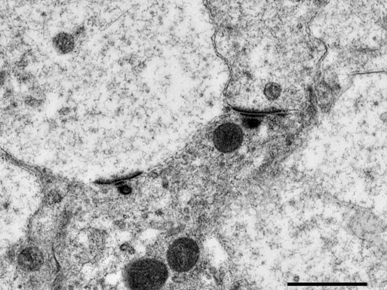
(Inner hair cell ribbon synapses in cochlear tissue)
TEM allows the resolution of subtle phenotypic differences within samples. (In collaboration with Steel.)

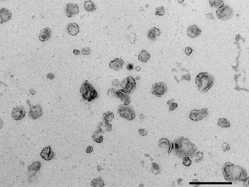
(Negatively stained exosomes)
TEM is used to characterise a nanovesicle preparation from cell culture supernatants of mesenchymal stem cells and assess the quality of the preparation method. (In collaboration with Filippi.)

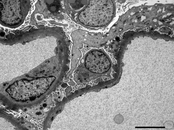
(Renal biopsy)
TEM is a powerful tool in the diagnosis of renal pathologies. In this case the sample was studied in relation to membraneous glomerulonephritis. (In collaboration with Guys and St Thomas’s NHS Foundation Trust.)
