Cryogenic Scanning Electron Microscopy (Cryo-SEM)
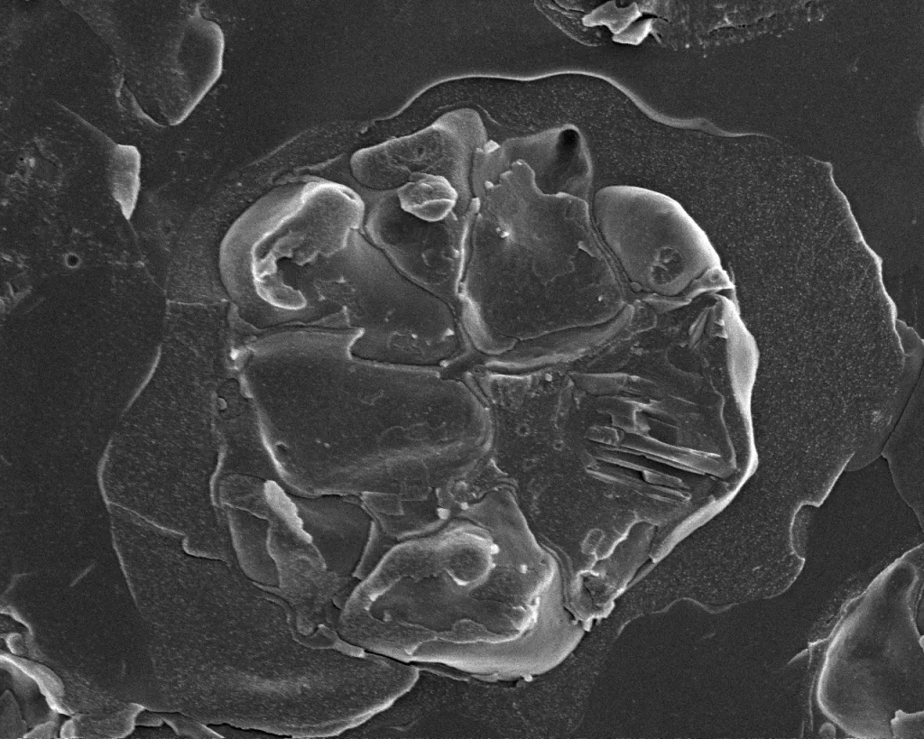
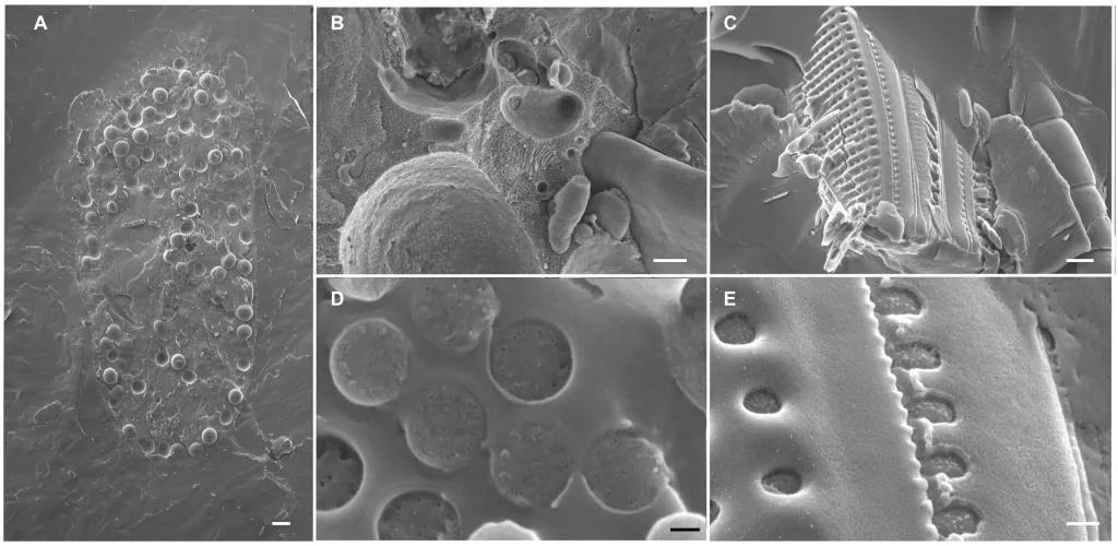
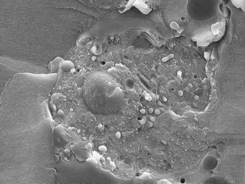
Cryo SEM is very useful for studying cellular and structural biology. This method allows research with sub nanometre resolution imaging at very low operating voltage. This protects sensitive samples while also maintaining the contrast and spatial resolution essential to understanding the key functions and processes of organelles.
We support the complete specialist workflow necessary to support high resolution cutting-edge cryo SEM. This allows direct investigation of membrane structure, subcellular organisation and cell-cell dynamics.
Modern SEM systems (with improved detector sensitivity and signal-to-noise) now achieve sub-nm resolution at 1 kV, making them highly capable tools for life science research, particularly when combined with high-resolution coating and fracture techniques.
Cryo SEM advantages:
- direct observation of biological structures in their native, cellular context with topographic information at the nanometer-scale
- no loss of ions/ molecules and direct observation of lipids
- maintains membrane permeability and organisation
- no osmotic effects
Examples of use
This technique is useful for the study of:
- lung tissue surfactants (components, distribution within alveoli, etc)
- membrane bilayers and protein distributions on membranes in Plasmodium falciparum infected erythrocytes
Equipment available
- JEOL JSM 7800 Prime with Leica-microsystems VCT500 cryo-stage


