This work is the result of an international collaboration requiring a broad spectrum of know-how embracing many disciplines ranging from math and fundamental physics to biology, chemistry, pathology, radiology and clinical patient management. Our team is in the lucky situation to have experts for each domain and to also work with funding bodies that shared our visions, supported us over the years, and allowed our ideas to turn into reality. Ultimately, the translation of this fundamental research to patients is also possible due to a close collaboration with our industrial partners.
Ralph Sinkus, Professor of Biomedical Engineering at the School of Biomedical Engineering & Imaging Sciences
31 July 2024
Researchers use waves to quantify blood vessels architecture
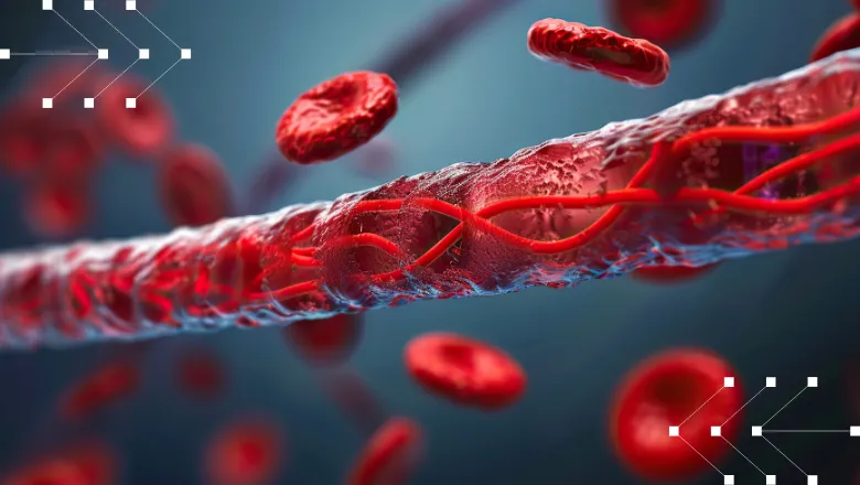
Researchers from the School of Biomedical Engineering & Imaging Sciences, along with partners at the University of Michigan, The Institut national de la santé et de la recherche médicale (Paris), Norway, and Germany are using shear waves to map blood vessel structures. Their goal is to improve treatments for tumours and other medical conditions.

The School's Professor Ralph Sinkus, and Professor David Nordsletten at the University of Michigan, have developed a new theory that uses MRI-based elastography imaging to study how shear waves travel through tissue. By analyzing these waves, they can measure the architecture of blood vessels non-invasively using readily available clinical imaging devices.
Shear waves carry important information about the materials they pass through, such as tissue stiffness, which can help diagnose diseases. The researchers aim to use this information to target drug delivery more effectively.
Their method relies on the dispersion of shear waves, which can reveal details about the microscopic structure of blood vessels. Dr. Sinkus explains that similar to how geophysicists use waves from earthquakes to study the Earth's interior, biomedical engineers can use MRI to observe waves inside the body. These waves can then be analyzed to understand the tissue's physical properties.
This method allows researchers to see tiny blood vessels that are usually too small to detect. Their theory and experiments show that blood vessels leave distinct signatures in the wave patterns, which can be detected and analyzed.
Dr. Sinkus uses an analogy to explain the concept: "Imagine trying to kick a football through a forest. If the trees are randomly scattered, the football will bounce around unpredictably. Similarly, waves travelling through tissue will be affected by the arrangement of blood vessels."
The team hopes that their research could significantly impact cancer treatment. Tumours often cause abnormal blood vessel growth, which is more chaotic than in healthy tissue. By measuring this chaotic structure, researchers can better understand tumours and potentially improve drug delivery. For instance, knowing how drugs travel through these vessels can help determine whether they reach the tumour cells or merely pass through without effect. This new approach could provide crucial insights into which drugs are best suited for different types of tumours, ultimately improving treatment outcomes.
Read the research paper now: Making sense of scattering: Seeing microstructure through shear waves


