Our team developed a very innovative solution to generate high-resolution 3D whole-heart images in a few minutes and in free-breathing. For the first time, a detailed depiction of the heart can be achieved in a clinically feasible acquisition time with MRI and with a high level of automation and efficiency, making this a nearly “push-button” solution for which we expect rapid clinical adoption.
Professor René Botnar and Professor Claudia Prieto, Professors of Biomedical Engineering, School of Biomedical Engineering and Imaging Sciences.
25 January 2024
Novel medical imaging technology enables detailed visualisation of the human heart
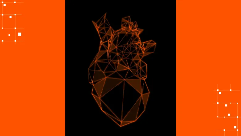
King’s researchers in collaboration with Siemens Healthineers have created a 3D visualization of the human heart via a novel technology based on Magnetic Resonance Imaging (MRI).

Professors Claudia Prieto and René Botnar and their King's academic teams at the School of Biomedical Engineering & Imaging Sciences (BMEIS) have developed a novel technological solution to provide 3D images of the whole human heart in an innovative, efficient, and patient-friendly way that is non-invasive.
The technology is based on new advanced MRI data acquisition, reconstruction and processing methods, developed in collaboration with local and global teams at Siemens Healthineers.
3D WholeHeart Pro is clear evidence of the value of collaboration of Siemens Healthineers on-site staff with our School’s academic research teams and R&D facilities. The opportunities created through this joint product development demonstrate how collaborative approaches to research can foster the rapid translation of novel medical technologies into clinical practice.
Professor Sebastien Ourselin FReng FMedSci, Head of the School of Biomedical Engineering & Imaging Sciences.
Cardiovascular diseases represent one of the leading causes of mortality across the world. MRI can help assess such diseases non-invasively by depicting thoracic vasculature (including the small and tortious coronary artery vessels), as well as evaluate myocardial tissue and cardiac viability. Conventionally, however, the routine acquisition of such magnetic resonance images in fine detail has been hampered by prolonged acquisition times and significant operator- and patient-dependency.
The newly developed framework is based on the under-sampled acquisition of MRI data, combined with a reconstruction algorithm that considers the deformations related to movements induced by respiration.
More traditional approaches would in fact require either the patient holding their breath or an inefficient rejection of imaging data in the case of inconsistent respiration in free-breathing. Differently, the designed approach enables the acquisition of sharp images without breath-holding even in individuals with irregular respiratory patterns, which are common in cardiac patients. In addition, this new technology simplifies MRI acquisitions by automatising several steps of the examination that would otherwise require manual interaction, thus improving usability and potentially limiting operator-dependent inaccuracies.
We are very proud to bring this novel technology into product. 3D WholeHeart Pro will allow clinicians to visualize the heart in three dimensions with high-resolution MR imaging. What is more, we expect the automated cardiac workflow to contribute to simplified operations in the hospital environment which in turn can increase access to MR imaging for more cardiac patients. Collaboration partnerships like the one at King’s College, where co-creation happens in synergy with our experienced on-site development team, are fundamental to innovation and to the advancement of technology – for the ultimate benefit of the patients.
Andreas Schneck, Head of Magnetic Resonance, Siemens Healthineers.
Research and development for MRI data acquisition and reconstruction methods primarily took place at St. Thomas Hospital in London in collaboration with the Siemens Healthineers on-site team (Karl Kunze and Radhouene Neji). At the same time, the novel automation features were developed by the Cardiovascular MRI R&D team at Siemens Healthineers Headquarters in Erlangen, Germany (Michaela Schmidt, Jens Wetzl, Seung Su Yoon). As part of this collaborative effort, the innovative framework was first deployed and tested at St. Thomas Hospital, leading to a large number of publications in peer-reviewed medical journals.
Following the first development stages in London, the solution was disseminated and validated at nearly 20 other institutions worldwide, including partners in the US, Germany, Switzerland, Denmark, Netherlands, China, Brazil, Hungary, Greece and others.
The developed technology is now being used as the basis of a product solution by Siemens Healthineers (*), global leader in medical imaging. The application, whose product name is 3D WholeHeart Pro, is being presented at the global Society for Cardiovascular MRI conference taking place in London (January 25th – 28th).
(*) The product is currently under development and not commercially available. Its future availability cannot be guaranteed.


