Accurate and timely treatment monitoring of breast cancer bone metastases is a recognised area of need and we hope that our programme of research into novel molecular imaging methods will improve management of individual patients by giving oncologists more accurate and timely information.
Prof. Gary Cook, Professor of Molecular Imaging, School of Biomedical Engineering & Imaging Sciences
20 March 2025
New Breast Cancer Imaging Trial underway at PET Centre
A clinical trial is underway to assess a novel radiotracer, [99mTc]-maraciclatide, which could enhance the early detection of breast cancer that has spread to the bones and provide quicker insights into treatment effectiveness.
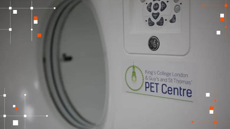
The trial, expected to run for approximately two years, focuses on how the tracer interacts with osteoclasts—bone cells that break down bone tissue. When breast cancer spreads to the bones, osteoclast activity increases, leading to weakened bones, fractures, and pain. By specifically targeting these cells, [99mTc]-maraciclatide may allow doctors to visually identify areas of bone destruction more accurately using SPECT/CT scans.
This new approach could significantly improve the detection of bone metastases and assess whether treatments—particularly those targeting osteoclasts—are working. If successful, the imaging technique may help clinicians determine much earlier whether a patient is responding to therapy, allowing for faster treatment adjustments and more personalised cancer care.
With further research and validation, this imaging breakthrough has the potential to revolutionise cancer treatment monitoring, offering hope to patients with advanced breast cancer in bones that is very difficult to monitor with standard scans.
The study is led by Prof. Gary Cook and Dr Amelia Moore. Participants have research SPECT/CT scans in the Nuclear Medicine Dept at Guy’s Hospital and PET scans in the King’s College London & Guy’s and St Thomas’ PET Centre.
Imaging patients whose cancer has spread to the skeleton is common reason for imaging and the Nuclear Medicine department sees a significant number of people for this very reason. Our department is pleased to be able to collaborate with Professor Gary Cook’s team to enable this novel molecular imaging research and for our Clinical Technologists, Radiographers and the rest of the team to contribute to this valuable work and help improve outcomes for breast cancer patients.
Damion Bailey, Education, Workforce & Professional Development Lead, Nuclear Medicine, Guy’s and St Thomas’ NHS Foundation Trust
The trial, which is a collaboration with the breast oncology team at Guy’s and St Thomas’ Hospital (GSTT) and the GSTT Nuclear Medicine Department, is supported by Serac Healthcare, who have supplied the tracer and is funded by Breast Cancer Now.
In this story

Professor of Molecular Imaging
Research Fellow in Osteoporosis