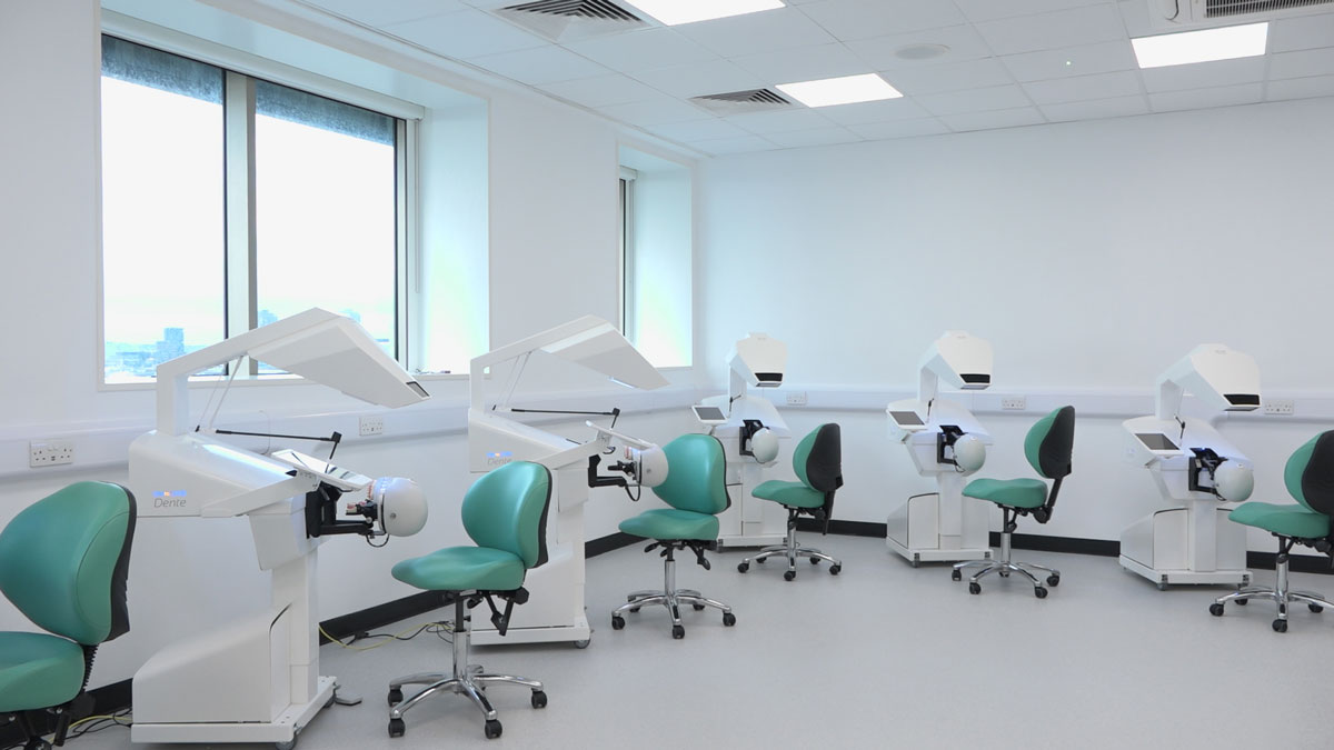
The Dental Simulation Fidelity Labs (FL18-A) are equipped with digital technologies including SIMtoCARE simulators and AV facilities, designed for a mixed augmented reality learning experience. Teachers can provide demonstration using one of the simulators projected on the screen, or seamlessly connect to a student’s simulator to show example practice exercises performed by learners. Students and teachers have a rich variety of learning experiences such as hands-on practice, tutorials and demonstrations.

Parts and functionality of the Simtocare Augmented Reality Simulator
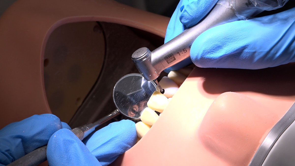
- Haptic device with real handpiece and mirror handle attachment supports operation using two hands simulating actual procedures e.g. drilling.
- The Phantom-head attachment with handpiece realistically simulates real-world practice.
- The 3D display screen allows the user to view the virtual mouth co-located within the manequin jaw where students can realistically experience drilling through the haptic device.
- An iPad displays the applications and treatments learners are performing allowing teachers to check on their progress and give feedback.
- Other features such as a foot pedal, height adjustment, and dental chair replicate clinical settings adding to the realism of the experience .
The Simtocare machine in operation
Dr Anitha Bartlett explains how the Simtocare high-fidelity haptic machines work.
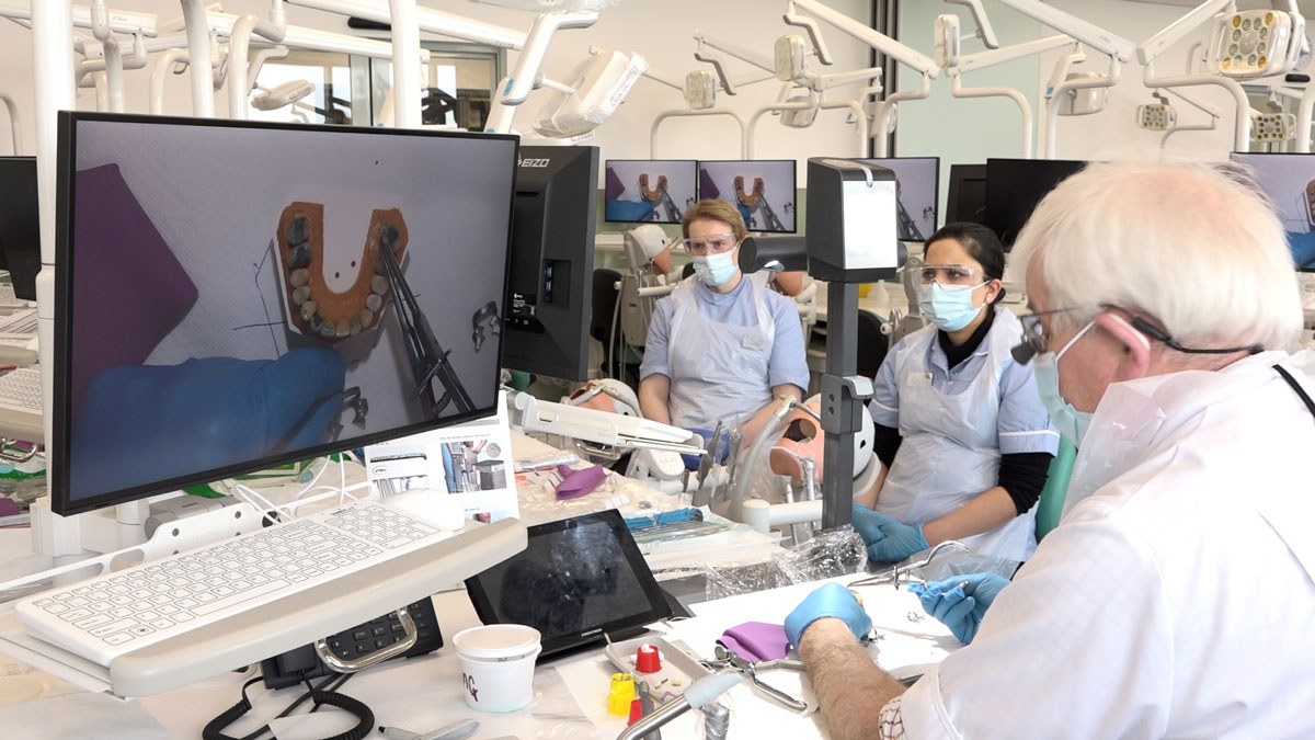
The Dental Simulation Fidelity Lab on Floor 20 of Guy's Hospital Tower Wing has the largest suite of dental CAD/CAM equipment in the world, with 10 intra-oral scanners and five milling machines. These are fully integrated with the 90 A-dec phantom heads in the facility, the FL18 AR simulators and the 275 dental chairs contained within the clinical facilities of Guy’s and St.Thomas NHS Foundation trust.
This means that real patients can be scanned in the dental chairs and their scans converted into AR for surgical practice in the FL18 simulators. The resulting surgical outcome (crown, bridge and implant preparations) can then enter the Planmeca CAD/CAM software on FL20 for design and milling of the dental prosthesis and the model of the real human anatomy to simulate and practice surgery before then carrying out the exact same treatment for real in the Guy’s and St. Thomas NHS Trust.
Access to this system means King’s students are uniquely equipped to experience and work within a truly integrated digitally augmented workflow. Students experience a complete delivery system that allows them to practice procedures develop skills, and receive meaningful feedback.
The FL20 facilitity is designed for blending lecture and lab to create an effective learning environment. While students are developing their skills, teachers can run demonstrations, provide feedback, and use digital modelling to help students understand real-world issues and how to approach them to ensure successful outcomes.
FL20 Fidelity Labs A, B, C and D.
Parts and functionality of the Adec Simulator
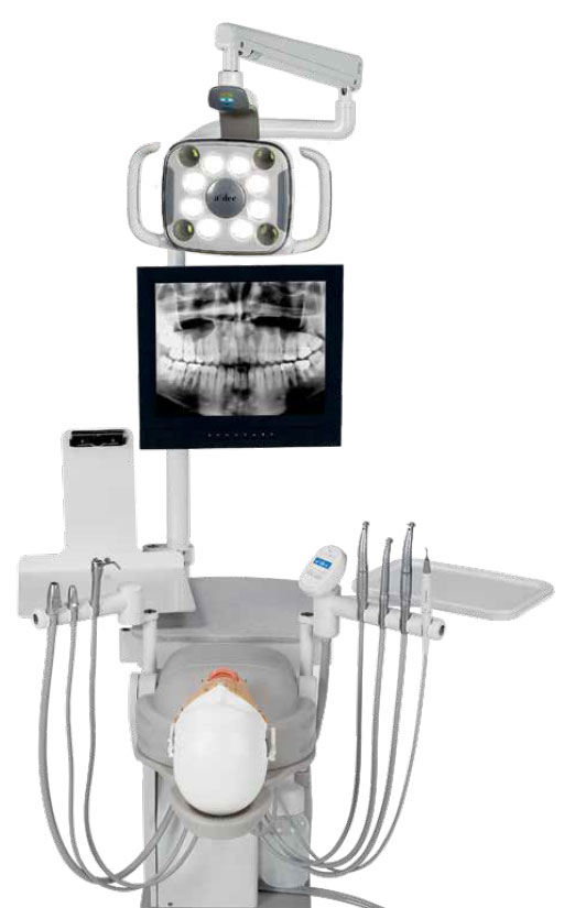
- Ergonomic setup – King’s is unique in allowing human teeth to be mounted into the simulator for clinical practice as close to real life as possible
- Manikin head – students operate and practice dental surgery with 1:1 anatomy
- Monitor display has multiple learning functions. Learners can watch teacher’s live demonstration from one of the simulators or work on ethically sourced simulated patient case[SDJ1] s which teachers present on the screen. In addition all student stations are enabled with CAD/CAM technology for 3D scanning, design and milling of state-of-the-art digital dental restorations
- Operation
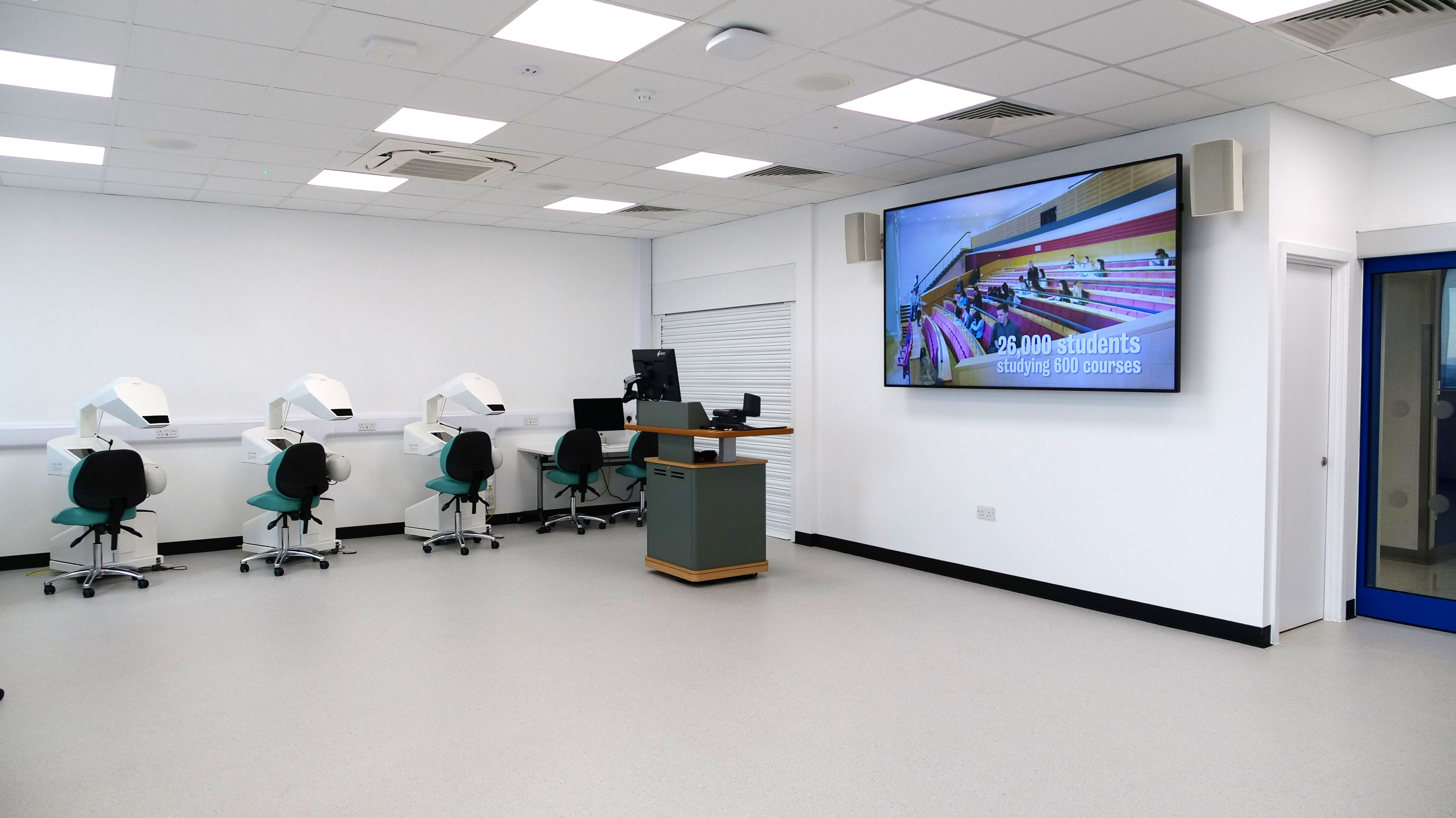 Each SimtoCare AR station is equipped with an iPad used to select training programmes and procedures, select appropriate tools and configure haptic settings. A visual representation of what the student sees is mirrored on the iPad so that teaching staff can monitor progress. It’s also possible for the visual output from any of the twelve AR stations to be sent to a large screen for demonstration purposes.
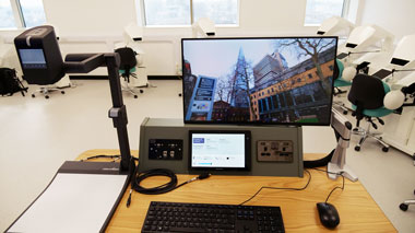
The Lecture capture system works across both the FL18 and FL20 labs, so joint sessions, for example incorporating an AR procedure in the FL18 lab using an intra-oral scan produced in the FL20 lab) can be seamlessly recorded.
Assessment of procedures completed by students can be reviewed by staff in-situ at the teaching station, which is also used to design further learning activities such as adding 3D scanned models from scans (see Fidelity Labs with ADEC). Other devices include a visualiser to allow teachers to project any analogue materials digitally on the screen.
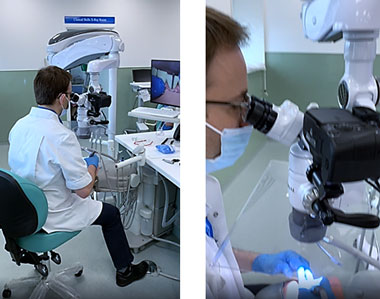
Training with surgical microscopes
Training with microscopes promotes better posture by reducing strain and fatigue while maintaining an upright sitting position when operating on simulated patients.The surgical microscopes have integrated beam-splitters to allow connection of a digital SLR camera for live-streaming dental surgery within the FL18 and FL20 labs and world-wide via the web.
The surgical microscopes allow minimally invasive precision surgery to be conducted at up to x35 magnification with a variety of light sources, including filtered for avoiding premature curing of photosensitive dental materials. These surgical microscopes allow learners to view details of dental surgery which it has not previously been possible to visualize, especially the intricate root canal anatomy of real human teeth.
From scans to haptics
Real patients are scanned in the dental chairs using intra-oral scanners and their scans converted into AR for surgical practice in the FL18 simulators. The resulting surgical outcome (crown, bridge and implant preparations) can enter the Planmeca CAD/CAM software on FL20 for design and milling of the dental prosthesis The exact same treatment can then be carried out for real in the Guy’s and St. Thomas NHS Trust. A facility of this size and integration is currently unique worldwide.
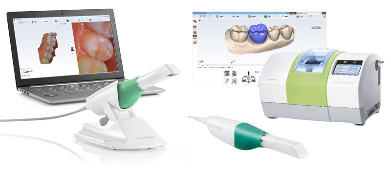
Emerald Intra-oral scanner photos courtesy of Planmeca Oy
This short video demonstrates an end-to-end digital workflow which begins with the creation of a 3D model of a patient's mouth using an intra-oral scanner. These scans can be loaded into the FL18 haptic sumulators, allowing students to gain experience of procedures using real-world data. The models are also used in the production of dental prostheses prior to real-life surgery.