New insight into the evolution of teeth
A team of researchers, led by King's College London Dental Institute, has provided new insight into the evolution and development of teeth.
Every jawed vertebrate needs teeth to function and feed, but exactly how teeth evolved is poorly understood because it is difficult to interpret different stages of tooth development from fossils. By looking at animals that build their teeth on a regular basis, like fish, scientists can begin to understand their development and arrangement in the mouth.
In a paper published today in Proceedings B, the Royal Society’s flagship biological research journal, the team demonstrates that two highly conserved dental genes, shh and bmp4 are differentially expressed in American paddlefish (Polyodon spathala). This fish is a member of the basal group of bony ray-finned fishes (Actinopterygii), presently the dominant group of vertebrates. Whilst over 30,000 species exist and have an immense range of dental morphological variation, the developmental regulation of the shh and bmp4 genes follows timing of classical stages of vertebrate tooth germ development.
Lead researcher Professor Moya Meredith Smith, from the Division of Craniofacial Development & Stem Cell Biology at the Dental Institute, said: ‘Our data shows design for expression of these genes is highly conserved amongst dentate jawed vertebrates, creating a template in a basal model animal for more comprehensive genetic regulation studies. As part of the NERC team, this genetic study is ongoing at Sheffield University. We will then know the genes that space the teeth out in a regular pattern, as in all dentitions, and maybe discover those for their regeneration.'
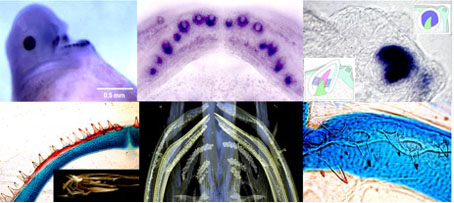 Morphogenesis of tooth development: embryo to larval to juvenile Polyodon.
The figure, top shows gene expression of shh at increasing magnifications: whole embryo in lateral view, shh localised expression in both upper and lower jaws; whole lower jaw in occlusal view, each +ve site of expression is a tooth germ formed at a slightly different time (spots are newest, rings later); close up of one at the cap stage, intense gene expression in cells around the tooth tip: bottom row at seven days old, tiny teeth are supported on cartilage (blue) of the jaws, CT scan of the juvenile with teeth on the jaws and on the gill arches of the pharynx (right, close up of pharyngeal teeth).
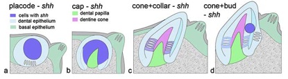
The four diagrams below explain the main stages through which all tooth germs go, with location of the active, or down-regulated gene expression in blue.
Notes to editors
'Making teeth to order: conserved genes reveal an ancient molecular pattern in paddlefish (Actinopterygii)' by Moya Meredith Smith et el. is published online in the Royal Society's journal Proceedings B on Wednesday 18 March 2015.